Post Mortem Contraction of Reissner’s Membrane and Meniere’s Disease
*Corresponding author: Daniel J. Pender, Department of Otolaryngology, Columbia University, United States of America. djp2djp2@gmail.com
-
Received: ,
Accepted: ,
How to cite this article: Pender DJ. Post Mortem Contraction of Reissner’s Membrane and Meniere’s Disease. Ann Otol Neurotol. 2025;6:e009. doi: 10.25259/AONO-2023-7-(201)
Abstract
Objectives
The degree and extent of endolymphatic hydrops in the cochlea are generally assessed by the displacement of Reissner’s membrane in the scala media. This has been graded as mild, moderate, and marked according to the positioning of Reissner’s membrane (RM). Any factors that influence the position of RM in the final temporal bone histology will have an impact on this assessment.
Material and Methods
The reported effects of elastic recoil, fixation contraction, decalcification, and dehydration shrinkage on Reissner’s membrane were surveyed, analyzed, and compared to a sampling of other tissues. These data were used to project the potential impact on the assessment of hydrops in the temporal bone histology.
Results
The assembled data on the effect of elastic recoil, fixation contraction, decalcification, and dehydration shrinkage suggest that Reissner’s membrane shortens approximately 15% due to post mortem tissue processing. Such a degree of shortening may be sufficient to reverse a mild to moderate displacement of Reissner’s membrane. It may also be sufficient to partially reverse a more marked displacement of Reissner’s membrane in the final histology.
Conclusion
Postmortem contraction of Reissner’s membrane due to processing effects is projected to be modest (~15% or less). This amount of membrane shrinkage could lead to a histological underestimation of the severity and extent of the hydrops that had existed during life and potentially obscure incipient Meniere’s disease.
Keywords
Endolymphatic hydrops
Meniere’s disease
Reissner’s membrane
Temporal bone
Tissue contraction
INTRODUCTION
Displacement of Reissner’s membrane (RM) in temporal bone histology (TBH) is the pathologic marker for endolymphatic hydrops (EH) in the cochlear duct. This has been graded as mild, moderate, and marked according to the positioning of RM.1 Any factors that influence the position of RM in the final TBH will have an impact on this assessment. However, the TBH presents only a final static endpoint image of RM. The degree and extent of RM’s displacement during life are not captured in the TBH, only the end result after post mortem tissue processing. If the displacement of RM in the living organism is substantially different from that seen in the TBH, then any diagnostic conclusions based on the TBH could be misleading, especially in connection with Meniere’s disease.
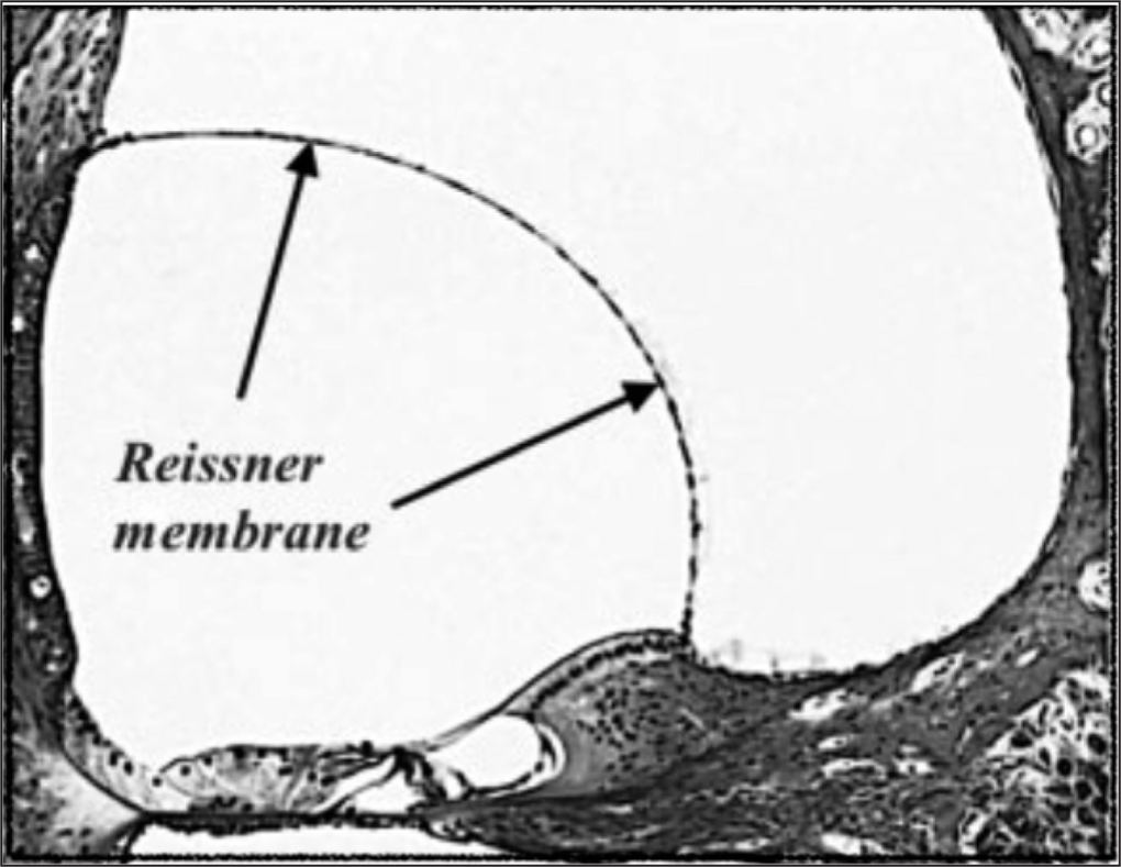
While assessments of RM displacements in-vivo can potentially utilize techniques such as radiology and surgery, assessments of RM displacement post mortem are usually confined to the final TBH. Several factors have been identified that, in the course of tissue processing, may contribute to a lessening in the final displacement of RM in TBH.2 Elastic recoil may occur in the interval after excision, and before the application of tissue fixative; fixation contraction may occur subsequently during preservative immersion; temporal bone decalcification then follows; and finally, tissue dehydration shrinkage may occur prior to hardening for microtome sectioning. The operation of these factors may lead to an endpoint assessment of the disease that differs in severity from that existing pre-mortem. However, the extent to which such factors affect the displacement of Reissner’s membrane is unclear. The object of this study was to assess the potential impact of these factors on the severity of endolymphatic hydrops as gauged by the histological displacement of RM, as shown in Figure 1.

- Reissner’s membrane displacement in endolymphatic hydrops.
MATERIAL AND METHODS
Reported effects on the various stages of post mortem processing of tissues were assessed and analyzed. This included elastic recoil, fixation contraction, decalcification, and dehydration shrinkage effects on RM and a sampling of other tissues. The composite effect on the degree of RM displacement was estimated from these data. This permitted a projection as to the potential impact on the interpretation of hydrops in the final TBH.
RESULTS
The impact of postmortem processing on RM displacement was reported according to the type of effect.
Elastic Recoil
Qualitative and quantitative assessments of elastic behavior by RM have been reported by several authors. Bekesy commented that RM cannot be dismissed as a slack tissue with negligible stiffness.3 Tonndorf reported a chance observation of RM viscoelastic behavior.4 Steele’s analysis deemed it permissible to model RM as a simple elastic material.5 Kimura reported that RM can exhibit purely elastic behavior over short intervals and more viscoelastic behavior in longer intervals.6 Elastic Recoil of RM was demonstrated in a hydropic guinea pig (GP) where the saccule had ruptured and thereby permitted RM to recoil to its normal position.7 Fresh postmortem human RM has been harvested and subjected to tenso-metric analysis.8 Such testing identified its distensile characteristics. A lower strain rate revealed a sigmoid viscoelastic pattern and a higher rate exhibited a rigid elastic pattern.9 Freshly excised GP RM has demonstrated 10% tissue recoil after excision.10
Fixation Contraction
Freshly excised Reissner’s membrane from guinea pigs is reported to exhibit an additional 5.9% contraction after immersion in 10% formalin. In contrast, a 15% contraction was noted with Heidenhain-Susa fixative, but only a 1–2% contraction with 3.1% glutaraldehyde.10
Decalcification Effect
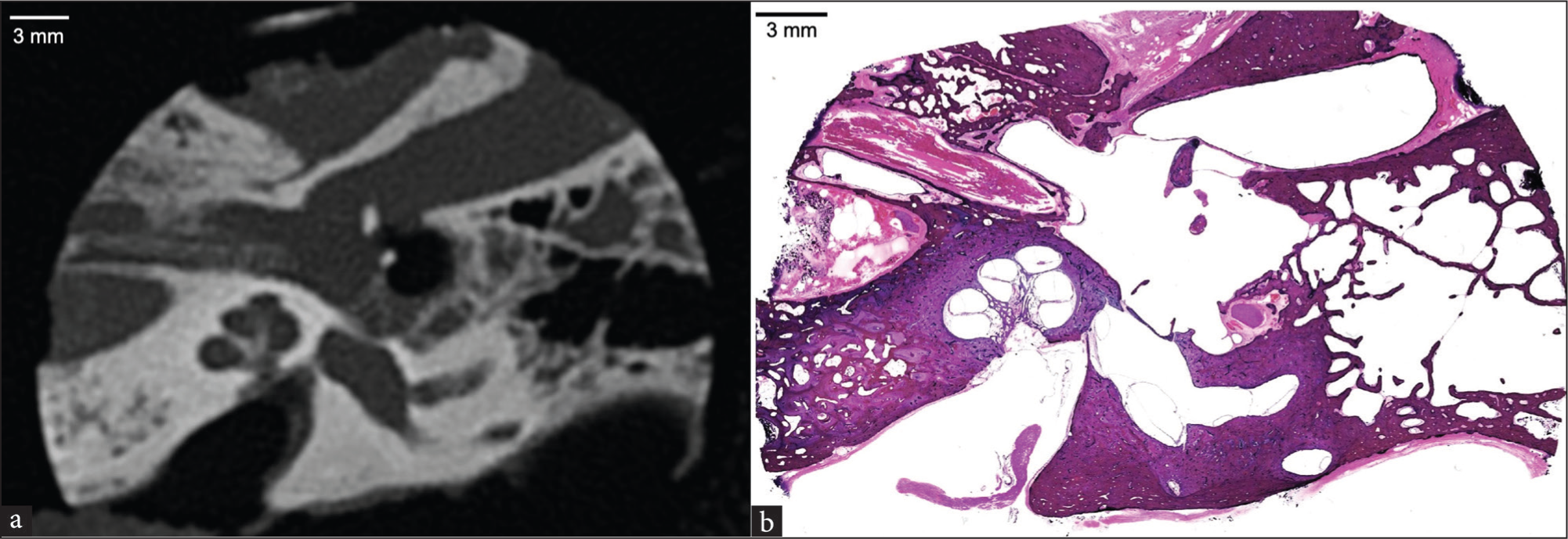
A comparison of computed tomography of a freshly excised temporal bone plug and the final TBH of the same specimen is shown in Figure 2.11 The bone plug computed tomography (CT) measures 34.4 mm in diameter, while the mounted histologic section measures 33.9 mm. This reflects a 1.5% overall specimen size reduction due to the removal of the calcium skeleton of the temporal bone plug.

- (a) Computed Tomographic image of temporal bone specimen before decalcification. (b) Same specimen after decalcification. Decalcification has resulted in a 1% reduction in measured size. (Courtesy of the Otopathology Laboratory at the Massachusetts Eye & Ear Infirmary).
Dehydration Shrinkage
Dehydration through alcohols is generally associated with tissue shrinkage.12 No specific data could be identified that addressed dehydration shrinkage of RM. However, human renal tissue was reported to shrink ~7% during histological processing2, and a sampling of excised ovine tissues, comprised of skeletal muscle, tongue, and oral mucosa, was reported to exhibit processing shrinkage averaging 19%13 [Table 1].
| Tissue type | Tissue source | Elastic recoil | Formalin contraction | Dehydration shrinkage | Overall size reduction | Reference |
|---|---|---|---|---|---|---|
| Renal | Human | 12% | 5% | 7% | 24% | Tran T, et al.[2] |
| Skin | Human | 20.7% | n/a | n/a | n/a | Kerns MJ.[17] |
| Skin | Human | 15.7% | 1% | n/a | n/a | Blasco-Morente G, et al.[18] |
| Labia | Ovine | 10% | n/a | n/a | n/a | Paramasivan S, et al.[13] |
| Muscle, mucosa, tongue | Ovine | n/a | 15% | 19% | n/a | Paramasivan S, et al.[13] |
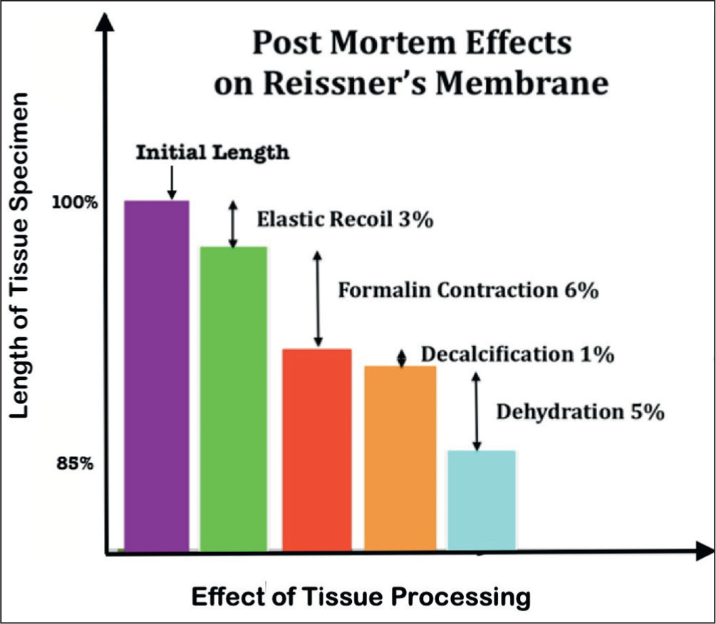
In sum, the combined shortening effects on Reissner’s membrane in the TBH can be conservatively estimated at 15% due to the contributions of elasticity of 3%, fixation of 6%, decalcification of 1%, and dehydration of 5%. These findings are summarized in Figure 3.

- Histogram of projected processing effects on the length of Reissner’s membrane.
DISCUSSION
Summary of the Results
Devitalized RM is estimated to exhibit an elastic recoil of 3%. Formalin fixation of RM is estimated to exert a contraction effect of 6%. Temporal bone decalcification is estimated to reduce specimen size by 1%. Dehydration shrinkage of RM is estimated at 5%. Total shortening of RM due to post mortem processing is estimated at 15%. This degree of RM shortening may be sufficient to potentially reverse mild to moderated degree of RM displacement and to partially reverse more marked degrees of displacement.
Analysis of the Results
Postmortem tissues appear to be subject in varying degrees to the influence of the ex vivo effects of elastic recoil, fixation contraction, and processing. This is most likely related to underlying differences in tissue molecular structure. Elastic tissue recoil is theorized to result from the reversal of molecular bond strain when trans-mural pressure abates post mortem due to the cessation of active metabolic processes. Fixation contraction is thought to result from formalin-induced hydroxy-methylene-based protein cross-linkage and coordinated calcium bonding.14 Decalcification effect is the result of the removal of the calcified skeleton of the temporal bone15 potentially allowing the remaining soft tissue to retract. Processing shrinkage during histological preparation appears to be the combined result of progressive alcohol dehydration, xylene adipose clearance, and celloidin embedding.16 Such tissue shrinkage results in specimen hardening that facilitates microtome sectioning. These effects occur in separately identifiable stages and, as such, have the potential to be individually quantified. In combination, they can exert a significant cumulative effect on TB specimens and thereby influence any anatomic appraisal of RM that relies on histology in terms of lesion appearance and relative dimension. This would imply that post mortem shortening of Reissner’s membrane, stretched as it is between the osseous spiral lamina and the bony wall of the cochlea duct, would result in a lessening of the displacement of RM and, therefore, a lesser degree of cochlear hydrops apparent in the final histology. This could alter the interpretation of cases of isolated apical hydrops as well as that of the contralateral ear in cases of Meniere’s disease and delayed endolymphatic hydrops.
Comparison with Published Data
No reports on the composite effect of elastic recoil, fixation contraction, decalcification effect, and dehydration shrinkage on RM post mortem could be identified. However, several studies were identified that reported processing effects in other tissues. For example, human skin was reported to exhibit elastic recoil of 15–20%,17,18 while demonstrating 1% fixation contraction. And ovine muscle, mucosa, and tongue specimens were reported to exhibit a 15% fixation contraction and 19% dehydration shrinkage.13 A study on renal tissue was the only comprehensive report that identified the original tissue size in vivo via imaging and then quantified the size reduction effect in each stage of processing, resulting in a cumulative reduction in length of 24% due to all factors.2 These are summarized in Table 1.
Limitations
The interior architecture of the temporal bone post mortem cannot be known with certainty before sectioning, given that the soft tissues contained within cannot be readily assessed in real time. This is especially true of Reissner’s membrane with its gossamer quality. Improvements in imaging may resolve this dilemma.
CONCLUSION
These cumulative data suggest that Reissner’s membrane very likely does exhibit all four post mortem tissue effects, namely elastic recoil, fixation contraction, decalcification effect, and dehydration shrinkage. These processing effects are projected to be modest (~15% or less) and may be sufficient to fully reverse and obscure a mild to modest displacement of RM while only lessening but not obscuring hydrops of a greater degree. Such an effect could alter the histological interpretation of isolated apical hydrops as well as our understanding of bilateral Meniere’s disease.
Ethical approval
Institutional Review Board approval is not required.
Declaration of patient consent
Patient’s consent not required as there are no patients in this study.
Financial support and sponsorship
Nil.
Conflicts of interest
There are no conflicts of interest.
Use of artificial intelligence (AI)-assisted technology for manuscript preparation
The authors confirm that there was no use of artificial intelligence (AI)-assisted technology for assisting in the writing or editing of the manuscript and no images were manipulated using AI.
REFERENCES
- Correcting the Shrinkage Effects of Formalin Fixation and Tissue Processing for Renal Tumors: Toward Standardization of Pathological Reporting of Tumor Size. J Cancer. 2015;6:759-766.
- [CrossRef] [PubMed] [PubMed Central] [Google Scholar]
- McGraw-Hill, New York: Experiments in Hearing; 1960.
- Endolymphatic Hydrops: Mechanical Causes of hearing loss. Arch Otorhinolaryngol. 1976;212:293-9.
- [CrossRef] [PubMed] [Google Scholar]
- Stiffness of Reissner’s Membrane. J Acoust Soc Am. 1974;56:1252-1257.
- [CrossRef] [PubMed] [Google Scholar]
- Experimental Blockage of the Endolymphatic Duct and Sac and Its Effect on the Inner Ear of the Guinea Pig. A Study on Endolymphatic Hydrops. Ann Otol Rhinol Laryngol. 1967;76:664-87.
- [CrossRef] [PubMed] [Google Scholar]
- Experimental Production of Endolymphatic Hydrops. Otolaryngologic Clinics of North America. 1968;1:457-47.
- [Google Scholar]
- Mechanical Properties of Human Round Window, Basilar and Reissner’s Membranes. Acta Otolaryngol Suppl. 1995;519:78-82.
- [CrossRef] [PubMed] [Google Scholar]
- The Distensibility of Reissner’s Membrane: A Comparative Analysis. Ann Otol Neurotol. 2022;5:21-27.
- [Google Scholar]
- Fixation-Induced Shrinkage of Reissner’s Membrane and Its Potential Influence in the Assessment of Endolymph Volume. Hearing Research. 1997;114:62-68.
- [Google Scholar]
- Available from:https://masseyeandear.org/assets/MEE/pdfs/ent/otopath/techniques-of-preparation-and-study-of-temporal-bones.pdf [Last accessed on 05.03.2023]
- Evaluation of the Temporal Relationship Between Formalin Submersion Time and Routine Tissue Processing on Resected Head and Neck Specimen size. Aust J Otolaryngol. 2021;4:32.
- [Google Scholar]
- Effect of Formalin Tissue Fixation and Processing on Immunohistochemistry. Am J Surg Pathol. 2000;24:1016-9.
- [CrossRef] [PubMed Central] [Google Scholar]
- Temporal Bone Normal Anatomy - Computed Tomography/ Histology Correlation. Otopathology Laboratory, Mass Eye and Ear Infirmary Available from:https://masseyeandear.org/assets/MEE/pdfs/ent/otopath/temporal-anatomy-05-2020.pdf [Last accessed on 05.11.2023]
- [Google Scholar]
- Shrinkage of Cutaneous Specimens: Formalin or Other Factors Involved. Journal of Cutaneous Pathology. 2008;35:1093-1096.
- [CrossRef] [PubMed] [Google Scholar]
- Study of Shrinkage of Cutaneous Surgical Specimens. J Cutan Pathol. 2015;42:253-257.
- [CrossRef] [PubMed] [Google Scholar]


