Translate this page into:
SmartNav in Cochlear Implantation: A Single Center Pilot Study
*Corresponding author: Bhawana Dangol, Department of ENT, Saifee Hospital, Mumbai, Maharashtra, India. drbdangol@gmail.com
-
Received: ,
Accepted: ,
How to cite this article: Dangol B, Kirtane MV, Satwalekar D. SmartNav in Cochlear Implantation: A Single Center Pilot Study. Ann Otol Neurotol. 2025;6:e008. doi: 10.25259/AONO_2_2025
Abstract
Objectives
The aim of this pilot study was to describe the feasibility and advantages of SmartNav in routine cochlear implantation as this was introduced for the first time in our setup. The objectives were to observe its usefulness to demonstrate speed, steadiness, and duration of electrode insertion along with impedance and neural responses. The study also explored the accuracy of the placement check run by software for the position of electrodes.
Material and Methods
The retrospective analysis of intraoperative and SmartNav records of all patients who underwent unilateral or bilateral cochlear implantation between Oct 1, 2024, and Dec 31, 2024, and evaluated with SmartNav system were carried out. Implantations were done in children and adults with profound hearing loss with normal inner ear anatomy as confirmed by imaging. A slim modiolar (CI632) or slim straight (CI622) electrode was used. The descriptive analysis of the parameters assessed by the SmartNav technology was carried out. The insertion parameters like speed, steadiness, and duration of insertion along with the electrophysiological parameters like impedance and neural response were analyzed. The accuracy of the placement check performed by the software was ascertained by comparing it with intraoperative C-arm imaging.
Results
The mean of the average speed and duration of insertion calculated in real time were 0.21 mm/sec and 1.4 minutes, respectively, for the CI632 electrode in 14 ears. These were 0.26 mm/sec and 2.35 minutes, respectively, for a case of CI622 electrode. The graphical depiction of the insertion process helped the surgeon to attain slow and steady insertion. SmartNav conducted simultaneous electrophysiological tests and correctly showed the electrode position in all cases.
Conclusion
SmartNav provides intraoperative feedback to the surgeon during the process of electrode insertion and offers the possibility of identifying the optimal speed for hearing preservation. It shows potential to replace intraoperative C-arm imaging for confirmation of correct array position, reducing the duration of anesthesia and radiation exposure. The electrophysiological monitoring performed by the software simultaneously with ongoing surgery saves operative time.
Keywords
C-arm
Cochlear implants
Electrode insertion
Electrophysiological test
Real time
INTRODUCTION
Cochlear implantation has revolutionized the management of profound hearing-impaired children and adults with successful outcomes. Apart from the surgery, electrophysiological tests are an integral part of the habilitation procedure. The tests comprising impedance and neural response telemetry (NRT) can evaluate the integrity of the device, placement of electrodes, and neural responses at the time of implantation and serve as a valuable tool to reassure the surgeon and family.1–3
The tests are performed in many centers along with the confirmation of electrode placement with intraoperative C-arm imaging and postoperative X-rays. Some surgeons do not conduct electrophysiological tests routinely with the notion that the surgical decision is rarely impacted with them and also because it requires extra surgical time.4 The evolution in the design of electrodes and electrophysiological tests has been improvised to detect tip fold overs (TFOs) commonly seen in thin and flexible perimodiolar electrodes.5
In 2022, CochlearTM released wireless real-time measurement technology called SmartNav. It evaluates real-time parameters like electrode insertion speed, angular depth of insertion, electrode placement check, and also impedance and NRT measurements. It acts like a navigation system as it feeds live information and provides feedback to the cochlear implant (CI) surgeon during the process of electrode insertion. The instant and ongoing feedback is of immense help that guides the surgeon for gentle, slow, and steady insertion of the electrode, minimizing the intracochlear trauma. This system has been described as user friendly, time saving, and efficient.6
This technology was introduced in our center and was used in all the patients with normal cochlear anatomy undergoing cochlear implantation with Nucleus® implants. As this was a novel approach for intraoperative measurements, we conducted a pilot study with an aim to describe its feasibility and advantages in routine cochlear implantation. The main objectives of our study were to observe its usefulness to demonstrate real-time parameters such as speed, steadiness, and duration of insertion of electrodes in addition to electrophysiological parameters like impedance and NRT. The study also explored the accuracy of the placement check run by the software to ensure the correct position of an electrode array.
MATERIAL AND METHODS
A retrospective chart review of the CI recipients operated and tested with the Cochlear™ Nucleus® SmartNav System in the period of 3 months from October 1, 2024 to December 31, 2024, was carried out as a pilot study. Both adults and children with profound hearing loss and with normal inner ear anatomy, as confirmed by imaging (HRCT scan and MRI), underwent either unilateral or bilateral implantation with Cochlear™ Nucleus® Profile™ Plus implants (CI622/CI632) at a private center for cochlear implantation. The study design complied with the principles stated in the Helsinki Declaration of 1975 (revised in 2000), and ethical approval for the study was taken from the Ethical Review Committee of the hospital.
The patients underwent preoperative audiological and radiological evaluation according to the standard CI protocol followed routinely to determine the candidacy for surgery. Cochlear SmartNav was used for testing intraoperative parameters and NRT.
All the implantations were performed under general anesthesia by the same CI surgeon. The surgical draping of the head was done with a thin sterile drape so as to ensure optimal distance between the surgical processor and implanted receiver/stimulator (RS). All the surgeries were done with the standard technique of cortical mastoidectomy/facial recess approach. Full exposure of the round window was carried out in all patients.
Atraumatic principles of electrode insertion were followed. Soft cochleostomy was done for CI632 slim perimodiolar electrodes to overcome angulation in the trajectory of insertion due to crista fenestra. The soft cochleostomy was created antero-inferior to the round window membrane for the placement of the electrode array in the scala tympani and to avoid trauma to the spiral ligament and basilar membrane.1 Round window insertion was done for CI622 slim straight electrode.
The RS was placed and fixed into the ramp created on the skull, followed by placement of the ground electrode into a sub-periosteal pocket underneath the temporalis muscle. The patient profile was entered in the SmartNav app on the iPad. Off-the-ear surgical processor covered in a sterile plastic bag was then hovered in a clockwise and anti-clockwise direction on the surgical drapes over the RS. This movement was continued till the processor adhered to the scalp area over RS with magnetic force, leading to the attainment of a stable wireless radiofrequency connection between RS and Custom Sound® Pro fitting software run on an iPad.
The SmartNav App version of 2.0.240300.137 was used, and the surgical processor used was CP 1150S. Live measurement of the parameters was initiated with the start of electrode insertion. The insertion speed was continuously measured and graphically shown in iPad screen. Continuous feedback was given by the system operator to the surgeon about the speed and steadiness of insertion as analyzed in the graph being obtained in real time. The angular depth of insertion was measured in a single patient implanted with a Nucleus Profile Plus CI622 slim straight electrode. The default measurement was computed for the diameter of the cochlea required for the measurement for the angular depth of insertion in this case. This measurement was not required in the case of implantation with the Nucleus Profile Plus CI632, as SmartNav does not measure the angular depth of insertion for pre-curved electrodes.
Real-time parameters like average speed and total duration of insertion were measured simultaneously during the process of electrode insertion. Post-insertion diagnostics were carried out after the completion of insertion. This comprised placement checks and electrophysiological tests like impedance and NRT. The normal placement of electrodes was shown in terms of green color coding of electrodes and the abnormal placement or potential TFO with red color coding of the affected electrodes. Similarly, the result of the impedance assessment was shown in green if normal and in red if it was a short or open circuit.
After completion of the electrophysiological testing, C-arm image was taken according to the routine protocol employed to check the electrode placement, as this was a pilot study. The wound was closed after the confirmation of correct placement. All patients were discharged within 24 hours. The electrophysiological data recorded in the SmartNav app were transferred to the appropriate center to facilitate further mapping sessions. The patients underwent a switch-on and mapping on the third postoperative week and further mapping sessions in subsequent weeks, adhering to the standard protocol.
The retrospective data on intraoperative electrophysiological measurements were computed into the data extraction sheet from the SmartNav records in the system. In the case of bilateral implantation, the parameters of each ear were entered separately into the data extraction sheet and analyzed independently.
RESULTS
SmartNav intraoperative measurements were conducted in 11 patients and 15 ears undergoing cochlear implantation in a period of 3 months from 1st October 2024 to 31st December 2024. Table 1 shows the demographic data of the patients included in the pilot study. Table 2 shows the findings of SmartNav in a total of 15 ears operated. Full insertion of the electrodes was attained in all implantations.
| Variables | Number/median/range | |
|---|---|---|
| Age (median; range) | 1.8 years; 1–54 years | |
| Gender | Male | 1 |
| Female | 10 | |
| Surgical approach to scala tympani | Round window | 1 |
| Cochleostomy | 14 | |
| Implants per patient | Unilateral | 7 |
| Bilateral | 4 | |
| Implant type | Nucleus CI622 slim straight | 1 |
| Nucleus CI632 slim modiolar | 14 | |
| CI: Cochlear implant. | ||
| SN | Age (years) | Implant type | Route of electrode placement | Average insertion speed (mm/sec) |
Insertion duration (minutes) | Placement check | Angular depth of insertion (degree) | Impedance results | NRT results (electrode number) | Array placement in C arm imaging |
|---|---|---|---|---|---|---|---|---|---|---|
| 1 | 7 | CI632 | Cochleostomy | 0.20 | 1.5 | Ok | – | Normal (1–22) | Present (1–22) | Ok |
| 2 | 2 | CI632 | Cochleostomy | 0.16 | 1.9 | Ok | – | Normal (1–22) | Present (1–22) | Ok |
| 3 | 2 | CI632 | Cochleostomy | 0.14 | 2.2 | Ok | – | Normal (1–22) | Present (1–22) | Ok |
| 4 | 6 | CI632 | Cochleostomy | 0.26 | 1.2 | Ok | – | Normal (1–22) | Present (1–22) | Ok |
| 5 | 1.8 | CI622 | Round window | 0.17 | 2.3 | Ok | 447 ±45 | Normal (1–22) | Present (1–22) | Ok |
| 6 | 1.3 | CI632 | Cochleostomy | 0.32 | 0.96 | Ok | – | Normal (1–22) | Present (1–22) | Ok |
| 7 | 1 | CI632 | Cochleostomy | 0.17 | 1.8 | Ok | – | Normal (1–22) | Present (1–22) | Ok |
| 8 | 1.7 | CI632 | Cochleostomy | 0.21 | 1.5 | Ok | – | Normal (1–22) | Present (1–22) | Ok |
| 9 | 1.5 | CI632 | Cochleostomy | 0.26 | 1.2 | Ok | – | Normal (1–22) | Present (1–22) | Ok |
| 10 | 1.8 | CI632 | Cochleostomy | 0.20 | 1.5 | Ok | – | Normal (1–22) | Present (1–22) | Ok |
| 11 | 1.8 | CI632 | Cochleostomy | 0.27 | 1.1 | Ok | – | Normal (1–22) | Present (2–22) | Ok |
| 12 | 54 | CI632 | Cochleostomy | 0.18 | 1.7 | Ok | – | Normal (1–22) | Present (2–22) | Ok |
| 13 | 1.6 | CI632 | Cochleostomy | 0.27 | 1.1 | Ok | – | Normal (1–22) | Present (1–22) | Ok |
| 14 | 1.6 | CI632 | Cochleostomy | 0.13 | 2.3 | Ok | – | Normal (1–22) | Present (1–22) | Ok |
| 15 | 9 | CI632 | Cochleostomy | 0.17 | 1.8 | Ok | – | Normal (1–22) | Present (1–22) | Ok |
| CI: Cochlear implant; NRT : Neural response telemetry. | ||||||||||
The mean of the average speed of insertion was 0.21 mm/sec for the CI632 electrode and was 0.26 mm/sec for the CI622 electrodes. The mean of the duration of insertion was 1.4 minutes for CI632 and 2.35 minutes for a case implanted with a CI622 electrode.
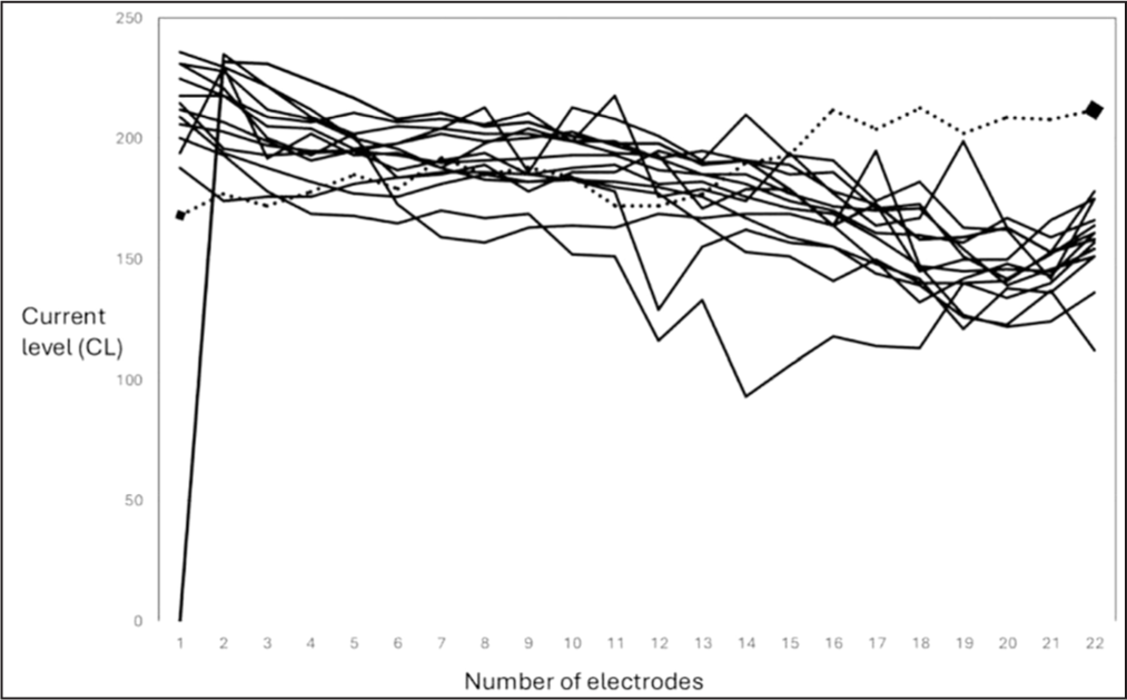
The NRT showed that current levels in apical electrodes were lower than the basal ones in all fourteen slim perimodiolar electrodes. One case implanted with a slim, straight electrode showed almost similar current levels in all electrodes. Figure 1 shows the line diagram of current levels in each electrode during NRT for a total cases of 14 perimodiolar and 1 straight electrode array.

- Line diagram showing current levels in 22 electrodes in total 15 implantations during neural response telemetry (NRT). Solid lines: Line diagrams showing current levels in 22 electrodes for CI632 slim modiolar electrodes; Dotted line: Line diagram showing current level in 22 electrodes for a CI622 slim straight electrode.
DISCUSSION
Insertion Speed and Steadiness
The average insertion speed ranged from 0.13 mm/sec to 0.32 mm/sec with a mean of 0.21 mm/sec for the CI632 electrode. In the single case of implantation with the CI622 electrode, the average speed was 0.26 mm/sec. The mean of the average speeds for CI632 electrodes and the average speed for CI622 electrode correspond to the recommendation of performing an insertion at a speed of around 0.25 mm/sec for hearing preservation.7,8 In a study on the application of SmartNav in cochlear implantation with CI632 electrodes, the mean of the average speed was 0.64 mm/sec.9 The literature has shown varied values of the speed of human insertions ranging from 0.7 mm/sec to 2.75 mm/sec in an experimental setting with contour advance electrodes and 0.25 mm/sec to 1 mm/sec in surgical implantations done with lateral wall electrodes.7,10 In one experimental insertion of lateral wall electrodes, the mean insertion speed attainable with the human hand was shown to be 0.87 mm/sec.11
The real-time graphical depiction of the insertion in terms of the speed and consistency is one of the major highlights of the application of this technology. The ongoing feedback obtained through the system operator focuses the surgeon for gentle, slow, and continuous forward insertion. Rajan et al. showed that slow insertion speed helped to attain complete insertions with less resistance and led to hearing preservations.7 During faster insertions, the non-compressibility of the perilymphatic fluid in the scala tympani leads to increased resistance and mechanical trauma on the basilar membrane and organ of Corti besides bending the electrode tip.7,12,13 An ability of the CI surgeon to perform continuous, slow insertion is the key factor for atraumatic electrode insertion.7,10,11 The steady insertion without stopping forward motion is regarded as beneficial to avoid the application of a larger force to overcome the static friction force inside cochlea.11 In a study on the insertion of electrodes in an experimental setup where movements were tracked with an optical tracking system, Kesler et al. found that the slowest speed was associated with the reversal of insertion with electrodes sliding back and forth with the possibility of more tissue damage.11
It is impossible to maintain constant speed during the entire insertion.7 The steady insertion can be attained with the use of robots, as the slow and steady insertion is said to be beyond human limits.11 However, these insertions lack haptic feedback, which is required to feel the resistance and to stop insertion according to the principle of soft surgical techniques.14 The steadiness of the insertion could be evaluated in real time through SmartNav technology for each insertion. The process of electrode insertion is otherwise a blind procedure performed by identifying the anatomical landmarks and guided by some tactile feedback. The monitoring and recording of both the speed and steadiness of insertion might be helpful in identifying the optimal speed within limits of human fine movements needed for hearing preservation.
Duration of Insertion
The duration of insertion ranged from 57.5 seconds to 2.35 minutes with an average of 1.4 minutes for full insertion of the CI632 electrode of 18.4 mm. The single case implanted with CI622 with a length of 24 mm took 2.35 minutes for complete insertion. The duration of insertion of 30 seconds or more is shown to be associated with hearing preservation.7 Much slower insertions with a duration of 1–2 minutes are also described in the literature.15,16
Angular Depth of Insertion
Angular depth of insertion was analyzed by SmartNav software in one implant with a slim straight electrode. The angular depth of insertion of 447 ± 45° obtained correlates with the deeper depth of insertion described for the lateral wall electrodes.17 The angular depth of insertion accounts for the variation in linear depth of insertion according to the size of the cochlea.17 As there is a positive correlation between the angular depth of insertion and speech perception performance, this parameter can be taken into consideration in predicting the postoperative outcome of the implantation.17,18
Placement Check
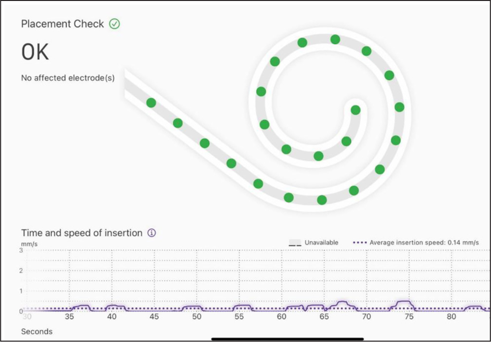
All the cases had correct placement of electrodes as shown in both SmartNav system and intraoperative C-arm fluoroscopy imaging. The placement check is essential for which imaging is considered the gold standard.19 The incidences of suboptimal electrode insertions like kinking, partial insertion, and TFO can occur and can lead to poor performances and facial nerve stimulations. This necessitates reimplantation when not identified and corrected intraoperatively. In our study, we used either slim perimodiolar or slim straight electrodes. The occurrence of TFO is common with perimodiolar electrodes with an incidence of 2–8% as described in the literature.20–22 Intraoperative imaging is necessary in such insertions as routine intraoperative electrophysiological tests to measure impedance and NRT often do not detect them.22 However, the SmartNav system can detect TFOs with the use of a TFO detection algorithm called the transimpedance matrix (TIM) algorithm.5 This is beneficial in situations when TFO can’t be properly detected by X-ray or CT scan in cases of blurring of electrode contacts due to an artefact.21 In a prospective study by Klabbers et al., all the TFOs were correctly identified by the TIM algorithm.19 The instant placement check performed with a built-in tool of TIM in SmartNav can be carried out simultaneously with ongoing surgical steps following electrode placement. This placement check reassures both the surgeon and patient, prevents the need for revision surgery, and has a potential to avoid C-arm imaging, preventing radiation exposure. Our results of 100% normal placement of array are in cases with normal cochlear anatomy only. The placement check is carried out by recording current levels in the electrodes as each one of them is stimulated. The flow of current in normal cochlea follows a specific pattern, unlike in an abnormal cochlea. The accuracy of SmartNav for the detection of abnormal placement like TFO can’t be commented on from our study as we did not encounter such incidence. However, the study by Kelsal et al. in 2022 showed the placement check algorithm with SmartNav was 100% accurate and specific in the detection of TFOs when compared with intraoperative imaging.6 Figure 2 gives a glimpse of the real-time graphical demonstration of electrode insertion and the placement check.

- Real time graph of electrode insertion and placement check run by SmartNav. The x axis of the graph represents time in seconds and y axis shows the speed of insertion in mm/sec. The dotted purple line shows average insertion speed which is 0.14 mm/sec for this insertion. The purple solid line depicts electrode insertion which is uniform.
Impedance
The impedances were normal for all the cases with the value less than 30 kΩ. There were no open or short circuits. The electrical impedance measurement checks device integrity. The instant confirmation of the normal functioning of the electrodes with the ongoing surgical procedure is a time saver for the surgeon.
Evoked Compound Action Potential
Thirteen out of 15 cases had evoked compound action potential (ECAP) in all electrodes in AutoNRT. Two cases did not have neural responses in the first apical electrode in Auto NRT as well as in Advanced NRT. Measurement of ECAP is a validated tool for setting an initial program for mapping.3,23 The attainment of neural responses in the form of ECAP is advantageous in cases of very young and uncooperative children that helps in the initial switch-on and mapping.1,3 The comparative analysis of the current levels in each electrode, as shown in Figure 1 during NRT in all implantations, shows the difference in the current levels in the apical electrodes in perimodiolar and straight electrodes. The lower current levels in apical electrodes in perimodiolar arrays are obvious due to their proximity to the modiolar wall and discrete spiral ganglion cell stimulation.24
The difficulty in maintaining a radio frequency (RF) link between the off-the-ear surgical processor and RS was only encountered in one adult due to a relatively thicker skin flap. This was dealt with transient manual pressure application and with the use of adhesives. The RF link maintained with such steps helped to complete SmartNav measurements. The correlation of age with the thickness of the skin flap is described with the range of 8 mm in adults in 3rd decade and 5 mm in the elderly in 9th decade.25 The maximum distance for magnetic locking of RS and the processor to create and maintain a connection is 7 mm.6 Hence, this issue was not encountered in children. However, thin, sterile surgical draping of the head is recommended to prevent this issue in all patients.
Constant communication between the surgical team and the SmartNav operator leads to efficient conduction of real-time diagnostics and electrophysiological monitoring. The use of the radiological placement check can become redundant with the use of this technology unless it is essential for the documentation and medicolegal purposes.
The SmartNav data is imported into Custom Sound Pro® fitting software, which can be retrieved for the MAP creation by audiologists involved in postoperative switch-on and mapping.
CONCLUSION
The initial experience on the application of SmartNav for the measurement of various intraoperative parameters during cochlear implantation was positive equally for the surgical team, operator, and audiologists involved in the switch-on and mapping. The technology is efficient and easy to use and time-saving to measure real-time and electrophysiological parameters. The real-time feedback possible with SmartNav guides the surgeon for slow and steady electrode insertion, which is one of the important factors of soft surgical techniques for hearing preservation. The accurate placement check run by the software has the potential to eliminate the need for intraoperative C-arm imaging to confirm the correct placement of the electrode array, thus reducing anesthesia duration and avoiding radiation exposure.
Ethical approval
The research/study approved by the Institutional Review Board at Saifee Hospital, Mumbai, Maharashtra, India, number SH/IRB/EXT/023/12/2024, 3rd January 2025.
Declaration of patient consent
Patient’s consent not required as patients identity is not disclosed or compromised.
Financial support and sponsorship
Nil.
Conflicts of interest
There are no conflicts of interest.
Use of artificial intelligence (AI)-assisted technology for manuscript preparation:
The authors confirm that there was no use of artificial intelligence (AI)-assisted technology for assisting in the writing or editing of the manuscript and no images were manipulated using AI.
REFERENCES
- How Well Do Cochlear Implant Intraoperative Impedance Measures Predict Postoperative Electrode Function? Otol Neurotol. 2013;34:239-44.
- [CrossRef] [PubMed] [PubMed Central] [Google Scholar]
- The Influence of Intraoperative Testing On Surgical Decision-Making During Cochlear Implantation. Otol Neurotol. 2017;38:1092-6.
- [CrossRef] [PubMed] [Google Scholar]
- Trends in Intraoperative Testing During Cochlear Implantation. Otol Neurotol. 2018;39:294-8.
- [CrossRef] [PubMed] [Google Scholar]
- Evaluation of a Transimpedance Matrix Algorithm to Detect Anomalous Cochlear Implant Electrode Position. Audiol Neurootol. 2022;27:347-55.
- [CrossRef] [PubMed] [Google Scholar]
- Early Clinical Experience with the CochlearTM Nucleus´ SmartNav System: Real-Time Surgical Insights [Internet]. 2022 [accessed 2025 Jan 20]. Available from: https://escucharahoraysiempre.com/wp-content/uploads/2022/09/SmartNav-1.pdf
- [Google Scholar]
- The Effects of Insertion Speed on Inner Ear Function During Cochlear Implantation: A Comparison Study. Audiol Neurootol. 2013;18:17-22.
- [CrossRef] [PubMed] [Google Scholar]
- Intracochlear Fluid Pressure Changes Related to the Insertional Speed of A CI Electrode. Biomed Res Int. 2014;2014:507241.
- [CrossRef] [PubMed] [PubMed Central] [Google Scholar]
- Intraoperative Measurement of Insertion Speed in Cochlear Implant Surgery: A Preliminary Experience with Cochlear SmartNav. Audiol Res.. 2024;14:227-38.
- [Google Scholar]
- Impact of the Insertion Speed of Cochlear Implant Electrodes on the Insertion Forces. Otol Neurotol. 2011;32:565-70.
- [CrossRef] [PubMed] [Google Scholar]
- Human Kinematics of Cochlear Implant Surgery: An Investigation of Insertion Micro-Motions and Speed Limitations. Otolaryngol Head Neck Surg. 2017;157:493-8.
- [CrossRef] [PubMed] [Google Scholar]
- A Model for Cochlear Implant Electrode Insertion and Force Evaluation: Results with a New Electrode Design and Insertion Technique. Laryngoscope. 2005;115:1325-39.
- [CrossRef] [PubMed] [Google Scholar]
- Force Application During Cochlear Implant Insertion: An Analysis for Improvement of Surgeon Technique. IEEE Trans Biomed Eng. 2007;54:1247-55.
- [CrossRef] [PubMed] [Google Scholar]
- An Optimized Robot-Based Technique for Cochlear Implantation to Reduce Array Insertion Trauma. Otolaryngol Head Neck Surg. 2018;159:900-7.
- [CrossRef] [PubMed] [Google Scholar]
- Hearing Preservation in Cochlear Implant Surgery. Int J Otolaryngol. 2014;2014:468515.
- [CrossRef] [PubMed] [PubMed Central] [Google Scholar]
- Hearing Preservation and Hearing Improvement After Reimplantation of Pediatric and Adult Patients with Partial Deafness: A Retrospective Case Series Review. Otol Neurotol. 2012;33:740-4.
- [CrossRef] [PubMed] [Google Scholar]
- Electrode Location and Angular Insertion Depth are Predictors of Audiologic Outcomes in Cochlear Implantation. Otol Neurotol. 2016;37:1016-23.
- [CrossRef] [PubMed] [PubMed Central] [Google Scholar]
- Influence of Cochlear Implant Insertion Depth on Performance: A Prospective Randomized trial. Otol Neurotol. 2014;35:1773-9.
- [CrossRef] [PubMed] [Google Scholar]
- Transimpedance Matrix (TIM) Measurement for the Detection of Intraoperative Electrode Tip Foldover Using the Slim Modiolar Electrode: A Proof Of Concept Study. Otol Neurotol. 2021;42:e124-9.
- [CrossRef] [PubMed] [Google Scholar]
- Development and Evaluation of the Modiolar Research Array—Multi-Centre Collaborative Study in Human Temporal Bones. Cochlear Implants Int. 2011;12:129-39.
- [CrossRef] [PubMed] [PubMed Central] [Google Scholar]
- Incidence for Tip Foldover During Cochlear Implantation. Otol Neurotol. 2018;39:1115-21.
- [CrossRef] [PubMed] [Google Scholar]
- Early Outcomes with a Slim, Modiolar Cochlear Implant Electrode Array. Otol Neurotol. 2018;39:e28-33.
- [CrossRef] [PubMed] [Google Scholar]
- Using Impedance Telemetry to Diagnose Cochlear Electrode History, Location, and Functionality. Ann Otol Rhino Laryngol Suppl. 1995;166:85-7.
- [Google Scholar]
- Surgical Experience and Early Outcomes with a Slim Perimodiolar Electrode. Otol Neurotol. 2019;40:e304-10.
- [CrossRef] [PubMed] [Google Scholar]
- Age-Dependent Variations of Scalp Thickness in the Area Designated for a Cochlear Implant Receiver Stimulator. Laryngosc Investig Otolaryngol. 2018;3:496-9.
- [CrossRef] [PubMed] [PubMed Central] [Google Scholar]


